Search Research Library:
Research Library
Publication
The Electricity of Touch: Detection and Measurement of Cardiac Energy Exchange Between People
Table of Contents:
3. Methods
Subjects were either seated in comfortable, high-back chairs to minimize postural changes or were lying down on a massage table. Prior to each session, subjects were informed of the tasks they were to perform and asked to refrain from talking, falling asleep or engaging in exaggerated body movements. The subjects were carefully monitored to ensure that there were no exaggerated respiratory or postural changes during the session.
Disposable silver/silver chloride electrodes were used for all bipolar ECG measurements. The positive electrode was located on the left side at the sixth rib and the reference was placed in the right supraclavicular fossa. Grass model 7P4 amplifiers were used for ECG amplification and Grass model P5 amplifiers were used for EEG and body surface measurements. The low frequency filters were set at the 1 Hertz setting and the high frequency filters at 35 Hertz. EEG electrodes were attached according to the International 10-20 system; the various recording sites and referencing are specified in each experimental session. Electrode resistance was measured with a UFI model 1089 electrode tester. Electrode to electrode resistance was typically in the range of 2 to 5 K. All data were digitized by a Bio Pac 16 bit digitizer and software system. The sample rate was 256 Hz. All post analysis was done with DADiSP/32 digital signal processing software.
All of the experiments monitored various recording sites on 2 subjects simultaneously. In all experiments, both subjects were wired with ECG electrodes as described above. To clarify the direction in which the signals were analyzed, the subject whose ECG R-wave peak was used as the signal time reference for the signal averaging is referred to as the "signal source," or simply "source." It should be emphasized that the subject designated as the source did not consciously intend to send or transmit a signal. The subject whose EEG or body surface recordings were analyzed for the registration of the source’s ECG signal is referred to as the "signal receiver," or simply "receiver." Signal averaging techniques were used to detect the appearance of the source’s ECG signal on the surface of the receiver’s body at various electrode locations. The resulting waveform appearing on the receiver is referred to as the signal-averaged waveform (SAW). The signal-averaged waveforms were triggered by the peak of the source’s ECG R-wave. The number of averages used in the majority of the experiments was 250 ECG cycles or roughly 4 minutes of data.

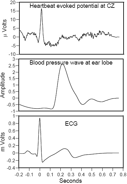
Figure 3.
Heartbeat Evoked Potential
Illustrates an example ofthe heartbeat evoked potential when a subject’s own ECG is usedas the signal source. The top trace is the EEG recorded at the CZlocation and the middle trace is the blood pressure wave, whichwas recorded at the earlobe. The signal from the heart arrives atthe CZ location around 10 milliseconds after the ECG R-waveand the blood pressure wave arrives around 240 millisecondslater.
It is well known that the electrical potential generated by one’s heartbeat can be recorded from any site on the body, including the sites recorded by the EEG.23, 26, 27 Figure 3 above. Therefore, in each of our experiments, the possibility had to be considered that the signal appearing in the receiving subject’s recordings was the receiver’s own ECG rather than that of the other subject designated as the source. Given the signal averaging procedure employed, this would only be possible if the ECG of the source was continually and precisely synchronized with the receiver’s ECG. To definitively rule out this unlikely possibility, in all experiments both the source and the receiver’s ECG were recorded.

